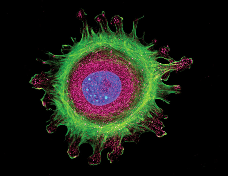
DIF is useful in the diagnosis of suspected autoimmune disease, connective tissue diseases and vasculitis. The staining patterns seen in tissue samples may be specific to a disease or may need to be interpreted with the clinical and histological findings.
What is Immunofluorescence?
01. Direct immunofluorescence
where antigens of interest have been bound in vivo by immunoglobulins with sampling of the skin.
02. Indirect immunofluorescence
where blood is taken to determine circulating antibodies to antigens of interest.
Direct immunofluorescence for the diagnosis of skin diseases
Autoimmune Bullous Diseases
- Pemphigus vulgaris
- Pemphigus foliaceus
- Paraneoplastic pemphigus
- Immunoglobulin (Ig) A pemphigus
- Bullous pemphigoid
- Pemphigoid gestationis
- Mucous membrane pemphigoid (cicatricial pemphigoid)
- Epidermolysis bullosa acquisita
- Bullous systemic lupus erythematosus (SLE)
- Linear IgA bullous disease
- Dermatitis herpetiformis
Vasculitis & Other Conditions
- Small vessel (hypersensitivity) vasculitis
- Henoch–Schönlein purpura
- Lichen planus
- Porphyria cutanea tarda
- Some adverse cutaneous reactions to drugs
- Photosensitivity rashes
Connective Tissue Diseases
- Lupus erythematosus (systemic, discoid, and subacute cutaneous forms)
- Neonatal lupus erythematosus
- Systemic sclerosis
- Mixed connective tissue disease
- Dermatomyositis
Where is the optimal site for biopsy?
The optimal site for taking a biopsy of the skin depends on the suspected disease.
Autoimmune Disorders
An optimal biopsy is a 3 or 4 mm punch biopsy taken from the perilesional skin (normal looking skin next to the rash) or mucous membrane tissue taken with tissue in concert with normal looking tissue.
Bullous Blisters
It is imperative that the active part of the rash or blister is taken with unreactive tissue so that a comparative analysis of tissue can be performed.
Vasculitis and Connective Tissue Diseases
A biopsy should be taken from lesional skin because uninvolved lesions are usually negative and in vasculitis, the reaction is evident in the vessels.
Transport and storage of the biopsy sample
Specimens should be placed in Michel’s fixative for transport.
Michel’s fixative can be stored at room temperature for up to two months.
Place a punch biopsy (preferable a 3-4mm punch) directly into the Michel’s transport media.
Place the container into a plastic bag and place the primary container into the shipping box at ambient temperature.
Ship the speciment to KorPath as soon as possible.


